Description
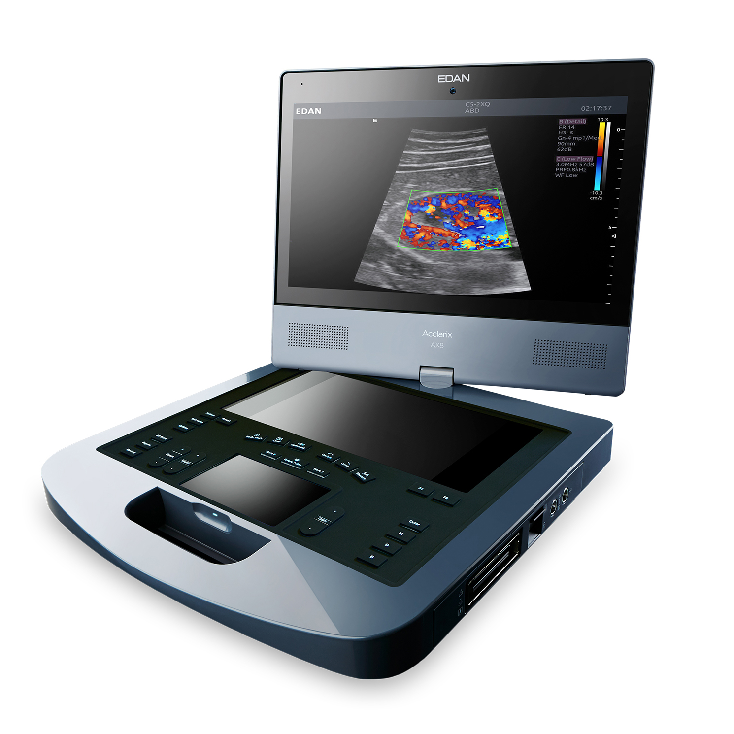
The Acclarix AX8 ultrasound system is a fully featured portable diagnostic ultrasound platform designed from the ground up with a relentless focus on delivering unexpected levels of innovation and performance. Born of a vision to deliver meaningful innovations that solve real clinical challenges the Acclarix AX8 features a distinctive design, definitive image quality, intelligent workflow and intrinsic quality.
Features:
- Tissue Adaptive Imaging
- Adaptive Doppler Imaging
- Needle visualization improves identification, even at steep angles
- Panorama
- Frequency compounding
- Spatial compounding
- Speckle reduction
- B-mode auto-optimization
User Interface
- Touch screen has three levels of access and drag-and-drop functionality for quick customization: Core functionality on one page, Swipe between pages for second tier controls, User created folders store infrequently used controls
- Unique dual touchscreen with second screen housing electronic virtual trackball: Gesture driven UI, Swipe to change, gain, scroll cine, Pinch in/out to zoom and resize color ROI
- Hard keys to access to core controls: Provides tactile feedback and landmarks for eyes-up navigation
- Sealed for easy cleaning
- Two programmable hard keys for direct access to most frequently used features
- Languages: English, Spanish, Ask about additional options
Environmental Operating Requirements
- Ambient temperature: 0° to 40°C
- Relative Humidity: 20%~80% (no condensation)
- Atmospheric pressure: 700hPa-1060hPa
- 110V-240V power supply
Cart Specifications (Optional)
- Snap-in anchoring of laptop into cart
- Height range of 31” to 40” to laptop palm rest
- Fixed deck angle of 15°
- Power converter housing built under tray
- Built-in tray houses printer and other incidentals
Connectivity
- DICOM: Verify SCP, Static image store SCU, Ultrasound multi-image store SCU, Four levels of compression, Data transfer options, Removable media, In-progress network storage, Auto-store at exam end, Manual-store on demand
- 4 USB ports (2 USB 2.0; 2 USB 3.0) Exports: DICOM studies, AVI and BMP files, PDF of report
- Video out: Display port and S-video
- Ethernet
Specifications:
System Architecture
- 128 channels
- Quad beam
- i7 processor with quad virtual cores
- 16Gb memory
- 500Gb hard drive storage
Mechanical Packaging
- Dimensions: W 38.8cm, D 40.7cm, H 7.7cm
- Weight: 9.25kg (includes battery)
- Main Screen: Tilt- and-swivel adjustment | 15” HD resolution (1920×1080)
- Magnetic monitor latch for secure transport
- Integrated handle provides wrist support during imaging
- Removable battery provides approximately 60 minutes of typical ultrasound use
- Optional multi-transducer connector
- Durable, ergonomic carry case
B-mode Imaging
- Tissue Adaptive Imaging provides continuous and automatic optimization including: dynamic range, speckle reduction, spatial compounding and persistence
- Enhanced border detection algorithms
- One-key auto optimization
- Digital zoom
- Depth: up to 30cm ( transducer dependent )
- Frequency range: Up to three fundamental and two harmonic frequencies per transducer
- Frequency compounding enhances penetration and detail
- Spatial compounding
- Speckle reduction
- Imaging formats: Curved, Linear, Phased array, Trapezoid, FOV for increased frame rate, Up/Down, Left/Right invert, Linear steered, Dual
- Additional optimization parameters: Gain and TGC, Dynamic range: 40-96, Map, Tint, Persistent, Focus position and number, Frame rate
Color Doppler
- Adaptive Doppler Imaging Automatically and continuously adapts to the flow state to optimize color fill-in, boundary detection and hemodynamic display.
- Supported modes: Velocity, Power Doppler Imaging (PDI), Directional PDI (DPDI)
- Side-by-side live format B-mode/color Doppler
- Additional optimization parameters: Gain, dynamic range, frame rate, frequency | Persistence, smoothing, wall filter, map | Steer angle, scale, invert, baseline, threshold
Strip Doppler
- Supported modes: PW, CW
- HPRF: Automatic invocation as needed to maintain gate location/scale
- Auto Doppler measurements: User selectable sensitivity and direction
- Duplex and Triplex displays
- Additional optimization parameters: Scale, Gain, Dynamic range, Wall Filter, Sweep speed, Baseline, Angle, Steer, Invert, Volume, Map and Tint, Frequency, Gate size, Strip size: Selectable top-bottom split screen display including full strip
M-mode
- Optimization parameters: Sweep speed, Persist, Map and tint, Strip size: Selectable top-bottom split screen display including full or side-by-side format, Gain, frequency, dynamic range (shared with B-mode)


Reviews
There are no reviews yet.